
Intranodular hyperechogenic figures - case 1473 (ultrasonographic picture 12)
doi: 10.24390/thyrocase1473ct.12
|
|

|
|
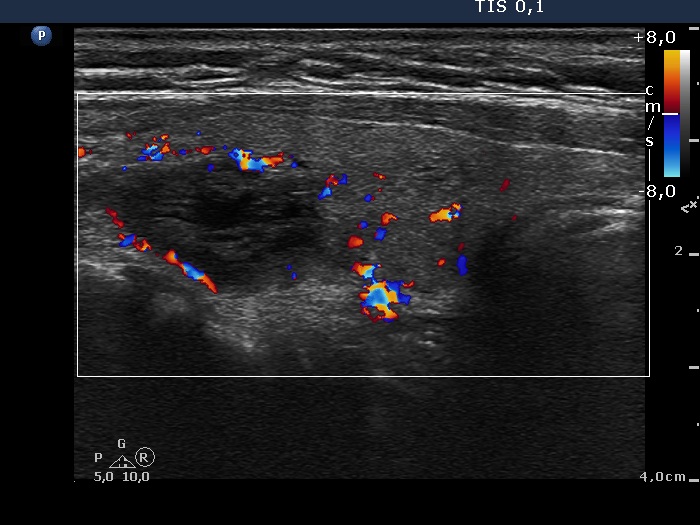
Right lobe, another longitudinal scan, color Doppler mode - after aspirating 9 mLcystic fluid. The nodule presents a type 2 vascular pattern, i.e. perinodular blood flow.