
Graves' disease - Case 23. (ultrasonographic picture 2)
|
|

|
|
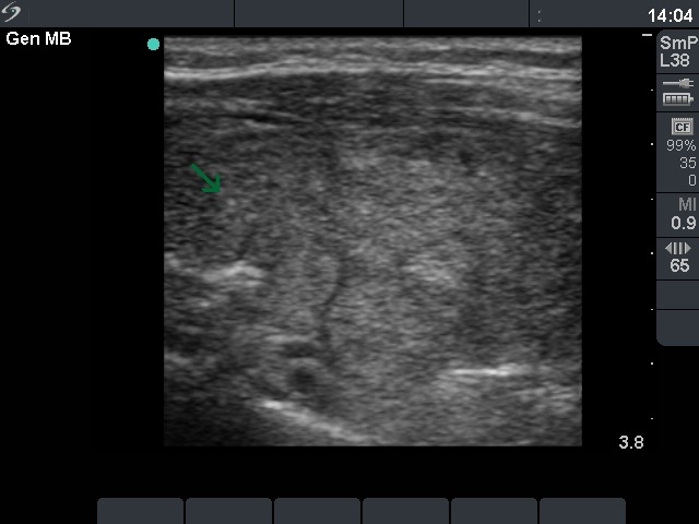
Right lobe, longitudinal scan. A moderately hypoechogenic lesion in the upper pole contains hyperechogenic punctate granulations. These may be foci of microcalcifications but more probably also that of fibrous tissues.