
|
|
Graves' disease - Case 23.
|
|


Clinical data: a 43-year-old man requested for an evaluation of hyperthyroidism.
Palpation: both lobes were enlarged and nodular.
Results of blood tests: hyperthyroidism with undetectable TSH-level. FT4 was 58.1 pM/L. TSAb-assay 12 U/L (normal value under 1.5).
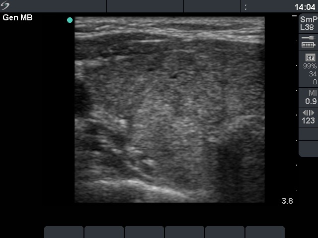
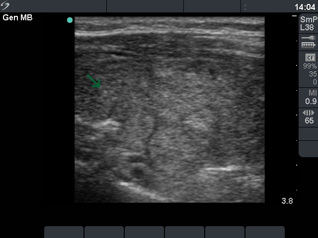
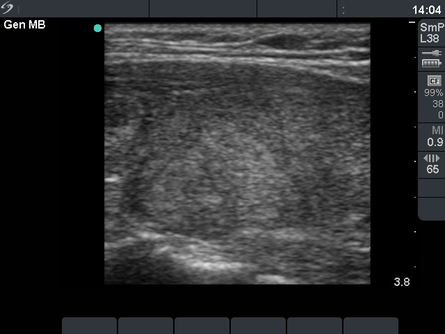
Ultrasonography: a moderately hypoechogenic thyroid was found with multiple circumscribed lesions divided by fibrous tissue. One of these areas contained hyperechogenic granules and exhibited increased vascularization.
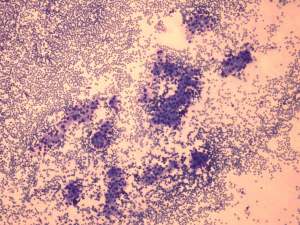
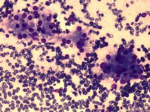
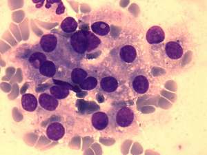
Cytological diagnosis: benign, corresponding to hyperthyroidism.
The patient was treated with thyrostatic drugs. After the normalization of FT4-level, he was operated on because of the size of his thyroid and the severe thyroid associated ophthalmopathy.
Histopathology: diffuse goiter.
Comments:
-
The US pattern of this patient highly resembles that of the so-called micronodular form of Hashimoto's thyroiditis. The "nodules" in this case are in truth pseudolobules of the lobes. The prominent appearance of the fibrous tissue dividing the lobules resembles a capsule of a follicular adenoma. In contrast with the latter, not only one or two, but numerous circumscribed areas are present in this case.
-
It is worth analyzing the so-called fire-flare appearance. This feature can be observed in smears stained by Wright-Giemsa staining.