
|
|
Graves' disease - Case 22.
|
|


Clinical presentation: a 36-year-old woman who was treated for hyperthyroidism for 2 years was referred now for an evaluation of a thyroid nodule discovered on ultrasound. Scintigraphy disclosed a "cold" nodule.
Palpation: a moderately firm nodule in the lower pole of the right thyroid.
Laboratory data: euthyroidism on daily 10 mg methimazole and 50 microgram levo-tiroxine (TSH 0.31 mIU/L, FT4 11.8 pM/L).
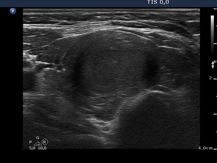
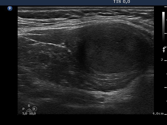
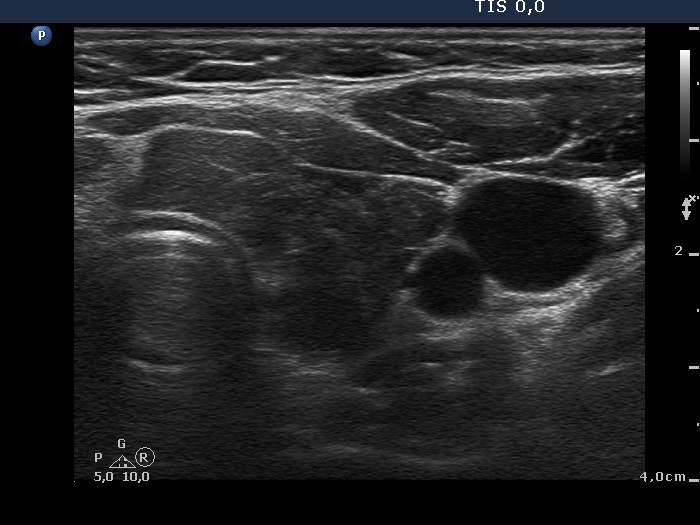
Ultrasonography: both thyroids were hypoechogenic and displayed fibrotic changes. There was a hypoechogenic nodule with blurred borders in the lower pole of the right lobe. The nodule presented a type 3 vascular pattern.
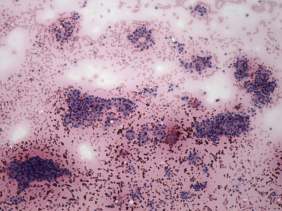
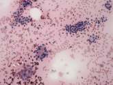
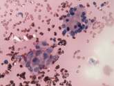
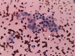
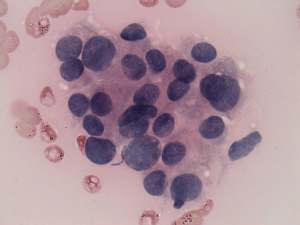
Cytology: besides groups corresponding to a benign goiter, there were a few groups of unusual, atypical, pleomorphic cells. Our diagnosis was the suspicion of papillary cancer.
Histopathology: disclosed benign hyperplastic nodules according to the lesion in the right lobe. Diffuse hyperplasia was found in the extranodular part of the thyroid.