
|
|
The operated thyroid - Case 5. doi: 10.24390/thyrocase1586.00Before and after total thyroidectomy - a patient operated on follicular carcinoma
|
|


First examination - before surgery (1st and 2nd rows of images)
Clinical data: A 21-year-old woman was referred for evaluation of elevated TSH-level detected on evaluation of fatigue.
Palpation: Both lobes were moderately firm.
Result of blood tests: subclinical hypothyroidism (TSH 4.19 mIU/L, FT4 13.8).
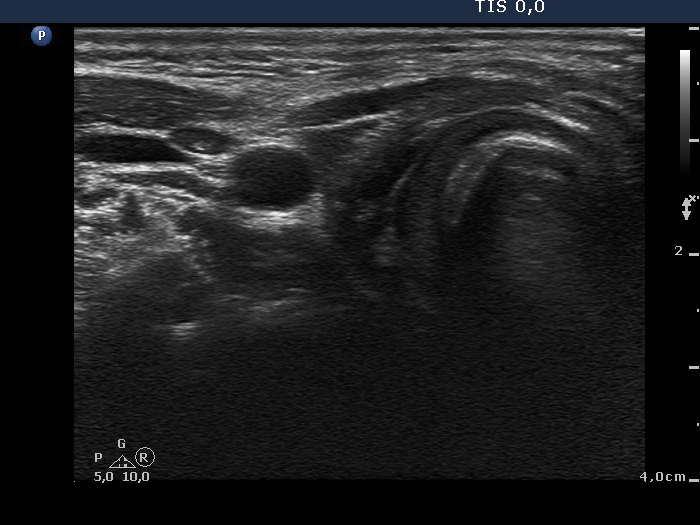
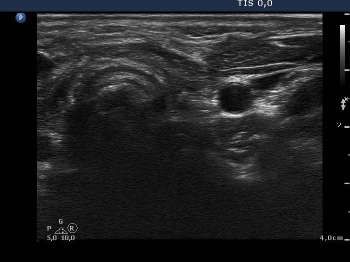
Ultrasonography. The thyroid was moderately hypoechogenic. There was a small nodule in the ventral part of the right lobe. It was hypoechogenic and presented microcalcifications. The vascularization was not specific.Aspiration cytology was performed from the nodule. Monolayered sheets predominated the smear, microfollicles were found, too. A few nuclei contained inclusions and grooves. The pattern itself was not sufficient to raise the suspicion of malignancy.
Taking the ultrasound presentation into account we gave a common ultrasound-cytological diagnosis of suspicion of papillary carcinoma and Hashimoto's thyroiditis.
Total thyroidectomy was performed. Histopathology disclosed Hashimoto's thyroiditis and follicular carcinoma with a maximal diameter of 3 mm according to the nodule. The tumor spread extracapsular.
Second examination - one year after surgery (3rd row of images)
Clinical data: The patient had no complaints.
Palpation: no abnormality.
Result of blood tests: euthyroidism on daily 100 microgram levothyroxine (TSH 2.89 mIU/L).
Ultrasonography: There was no parenchyma according to the thyroid which was replaced by connective tissue.
Comments.
-
To judge the presence of halo and perinodular blood flow is difficult in the event of a small lesion.
-
We presented the cytological smear in a postgraduate course. The opinion of experienced thyroid cytopathologist were divided almost equal as to whether the pattern is benign or belongs to atypia of undetermined significance or is suspicious for malignancy.