
|
|
Other rare thyroid tumors and non-thyroidal lesions in the region of the thyroid - Case 2.Poorly differentiated inzular carcinoma |
|


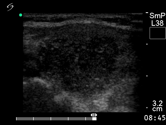

Clinical presentation: a 36-year-old woman was referred for an evaluation of a newly discovered nodule in the thyroid.
Palpation: a hard nodule was palpable in the right lobe.
Functional state: euthyroidism (TSH-level 2.09 mIU/L).
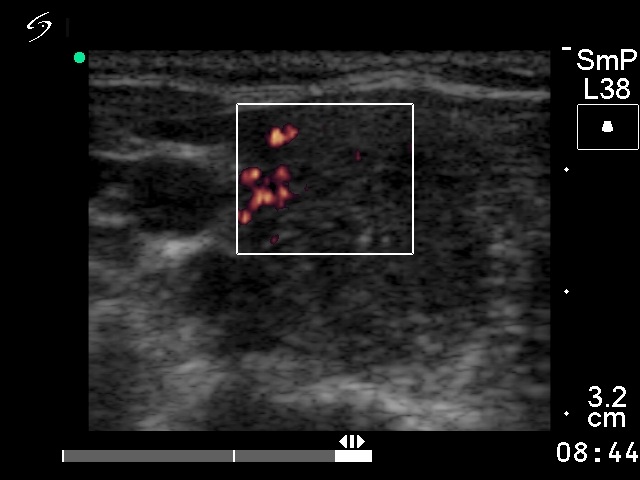
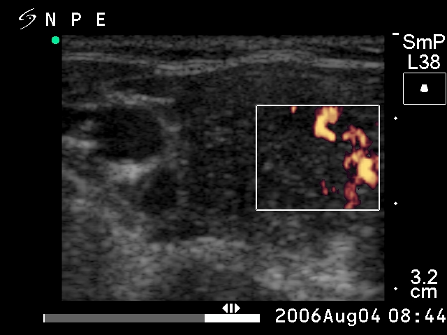
Ultrasonography: there were several hypoechogenic nodules within echonormal background. The dominant nodule was located in the right lobe. It was hypoechogenic, inhomogeneous. The borders of the nodule were irregular. The intranodular blood flow was increased.
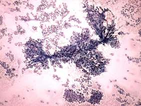
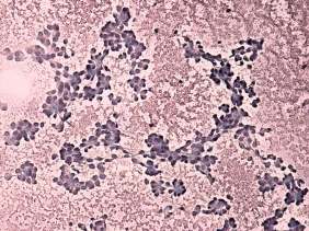
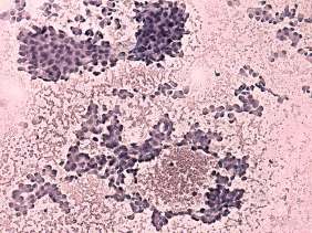
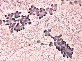
Cyological picture: there was no colloid in the background. Follicular cells occurred in microfollicles, in syncytial clusters and in arborizing structures with hyalinizing stroma. The single cells were polygonal and had eccentric nuclei. The nuclei were enlarged, several cells contained inclusion or groove. Cytological diagnosis: papillary cancer.
Histopathology: poorly differentiated inzular carcinoma.
Comments:
-
The correct interpretation of polygonal cells was difficult. The presence of polygonal cells could be suspicious for medullary cancer, but the structure of cells i.e. the predominance of microfollicular structure excluded this possibility.
-
The insular cancer has no specific feature. Both the cytological atypia and the architecture of cells varies.