
The role of complex diagnosis - follicular proliferation - Case 7. (cytologic picture 5)
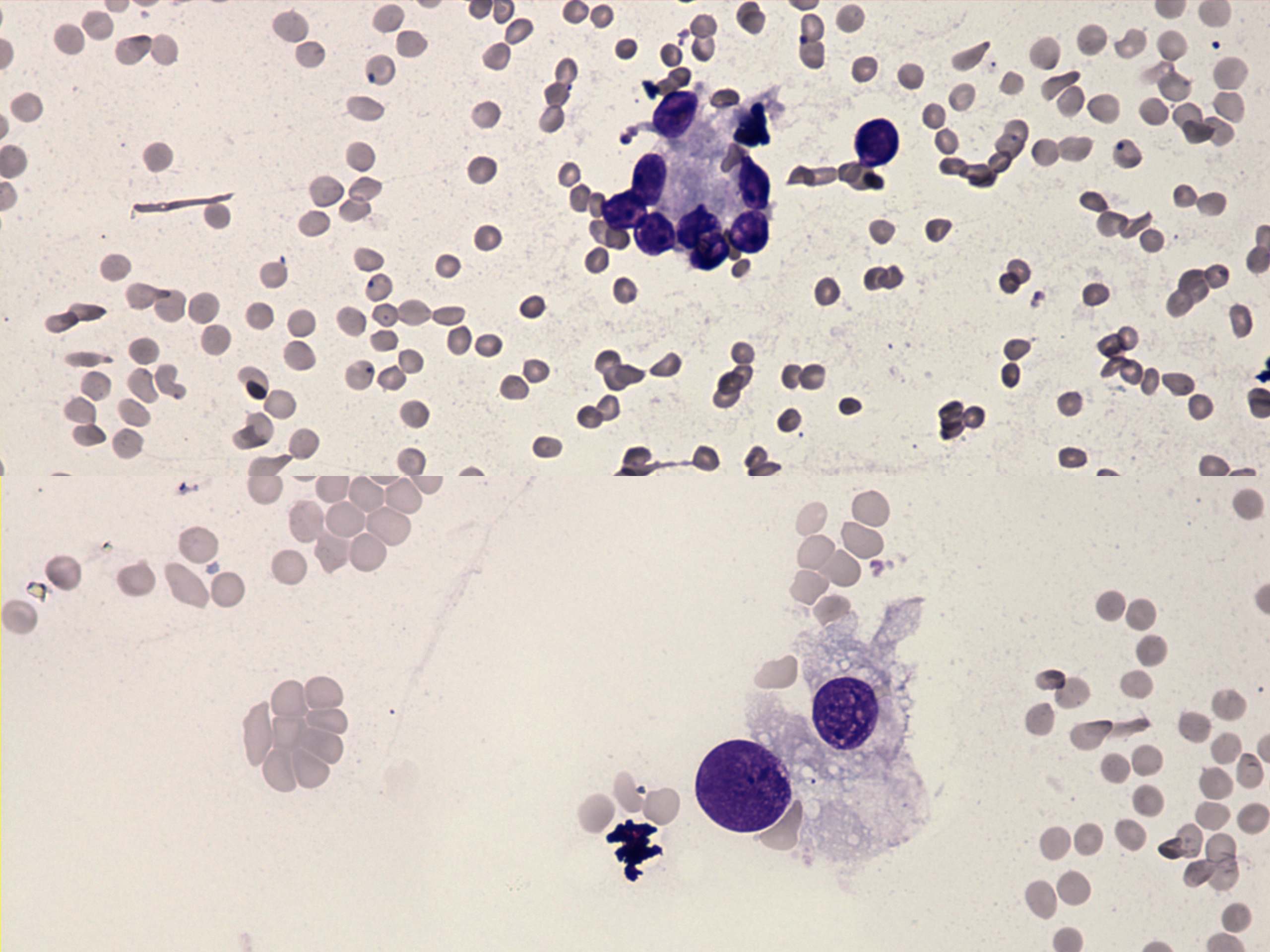 Wright-Giemsa staining, 400x. There is an irregular microfollicle in the upper part of the image displaying nuclear crowding. The two cells in the lower part present cytoplasmic vacuolization. |


