
|
|
An operated thyroid - Case 1.A patient with an unilateral subtotal thyroidectomy
|
|

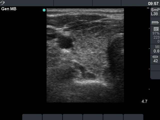
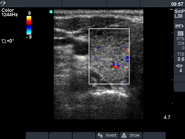
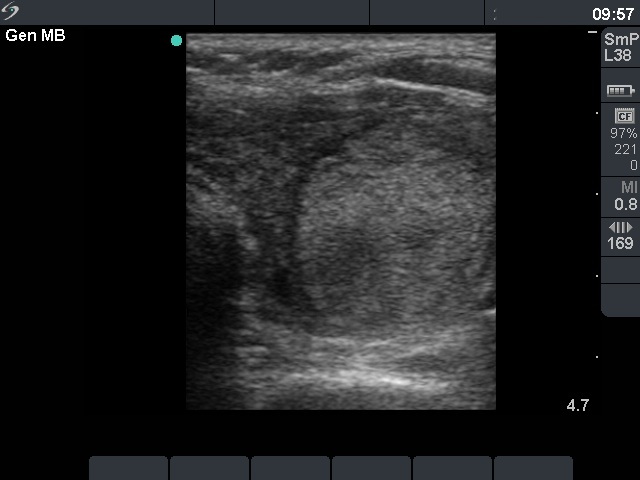


First examination - before surgery (1st row of images)
Clinical presentation: a 47-year-old woman was referred for evaluation of nodular goiter. She noticed a lump in the left lobe for a month.
Palpation: a large nodule in the left lobe.
Results of blood test: euthyroidism (TSH 3.31 mIU/L).
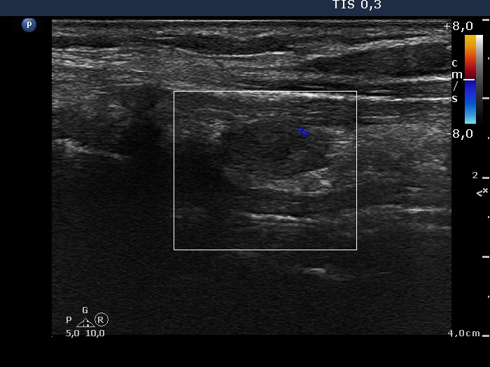
Ultrasonography: the right thyroid was echonormal and contained hypoechogenic areas. The left lobe was moderately hypoechogenic. There was a large hyperechogenic nodule in the left thyroid. The nodule displayed halo sign and type 2 vascular pattern.Cytology resulted in benign lesion.
Final diagnosis. Benign nodule in the left thyroid. Chronic lymphocytic thyroiditis. Left lobectomy was advised because of the size of the nodule.
Histopathology disclosed benign hyperplastic nodule and chronic lymphocytic thyroiditis.
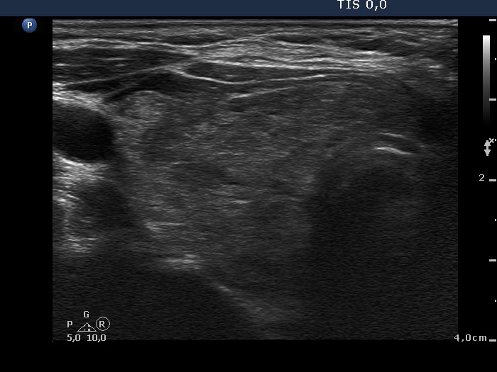
Second examination - 3 years after surgery (2nd row of images)
Comments.Clinical data: the thyroid function of the patient was regularly checked. She was euthyroid until the last blood test. She had no complaints except for a 5 kg gain in weight.
Palpation: the right thyroid was enlarged.
Results of blood tests: subclinical hypothyroidism (TSH 5.18 mU/L, FT4 12.7 pM/L).
Ultrasonography: the right thyroid was minimally-moderately hypoechogenic and enlarged and contained several hypoechogenic areas. The resected left lobe was moderately hypoechogenic.A replacement therapy with 50 microgram levothyroxin was started.
-
Before surgery the thyroids presented unequivocal signs of lymphocytic thyroiditis: the left thyroid was moderately hypoechogenic while the right displayed hypoechogenic spots within an echonormal background.
-
Three years after surgery the right thyroid became enlarged because it had to take over the hormon production of the resected left thyroid.