
|
|
Intranodular hyperechogenic figures - case 1726
|
|


Clinical presentation: a 37-year-old woman visited us for a follow-up of a nodular goiter. First she was investigated 4 years ago when a cystic nodule was found and 2 mL brown fluid was aspirated from the lesion. She had no complaints.
Palpation: the right lobe was suspicious containing a nodule.
Functional state: euthyroidism with TSH 1.60 mIU/L.
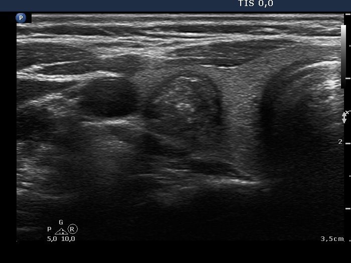
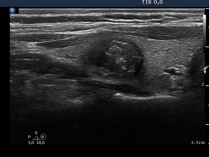
Ultrasonography. The thyroid was echonormal. There was a moderately hypoechogenic nodule. The lesion had small cystic areas and showed two forms of hyperechogenic intranodular granules; they partly were bright and corresponded to microcalcifications, partly belonged to the non-specific subgroup. The lesion presented signs of perinodular blood flow. Compared with the previous examination the nodule was smaller, 17x15x19 mm and 11x10x14 mm, first and current examination, respectively. It means that the cyst had not refilled in the past four years.
Aspiration cytology was performed because at the first examination only macrophages were on the smear. This time cytology resulted in benign cystic-colloid goiter.
Comments. The bright granules cannot be categorized other than microcalcifications.