
Intranodular hyperechogenic figures - case 1730 (ultrasonographic picture 2)
doi: 10.24390/thyrocase1730.02
|
|

|
|
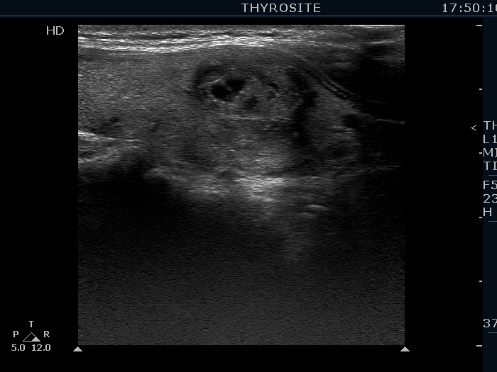
Right lobe, longitudinal view. The hyperechogenic lines dividing the lesion into smaller parts correspond to connective tissue as do the small granules.