
Differentiation of discrete lesions - case 1750 (ultrasonographic picture 8)
doi: 10.24390/thyrocase1750ln.08
|
|

|
|
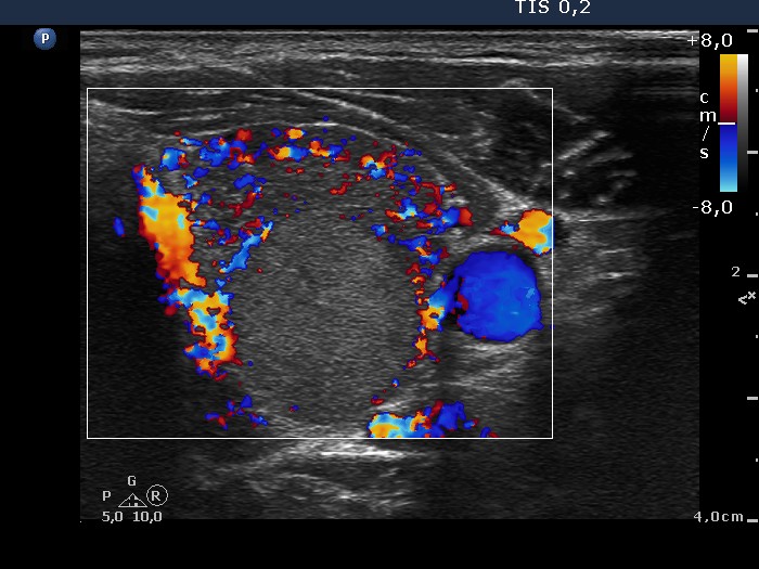
Left lobe, horizontal scan, color Doppler mode. This pattern is deceptive, the nodule in fact has neither perinodular nor intranodular blood flow; the extranodular part of the lobe presents markedly increased vascularization.