
Differentiation of discrete lesions - case 1750 (ultrasonographic picture 9)
doi: 10.24390/thyrocase1750ln.09
|
|

|
|
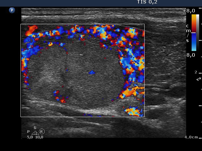
Left lobe, longitudinal scan, color Doppler mode. The nodule seems to have perinodular blood flow, while in reality only the extranodular part of the lobe presents markedly increased vascularization. The presence of a perinodular blood flow can be judged only on the dorsal part of the nodule which lacks circulation, therefore we can state that the nodule displays neither perinodular nor intranodular blood flow.