
Follicular adenoma - Case 21. (ultrasonographic picture 2)
|
|

|
|
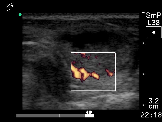
Left lobe, horizontal scan, power Doppler method. The lesion shows perinodular vascularity. The intranodular vascularization is detectable, but not increased.