
The role of complex diagnosis - follow-up of follicular lesions - Case 10.
5 years after initial investigation (ultrasonographic picture 3)

|

|

|
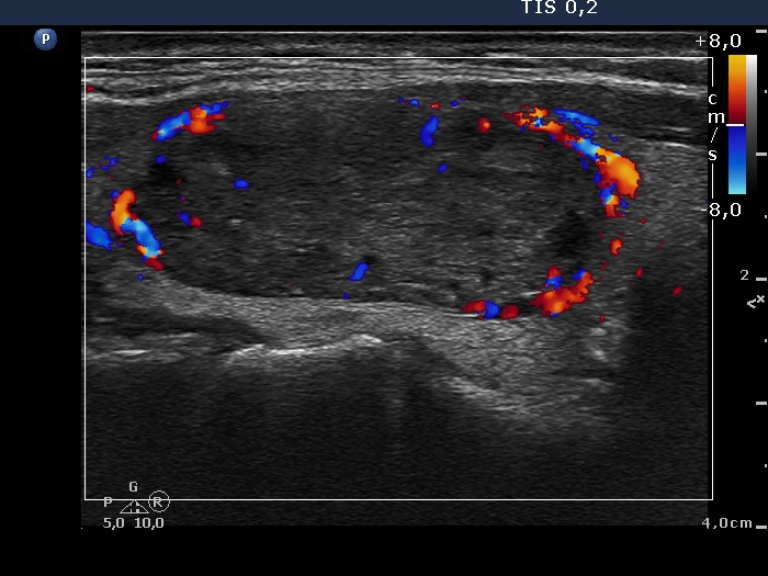
Right lobe, longitudinal scan, color Doppler mode. The lesion presents a type 2 vascular pattern with signs of a type 3 pattern, too.


