
The role of complex diagnosis - oxyphilic lesions - Case 2.
Two years after initial investigation (ultrasonographic picture 1)
|
|

|
|
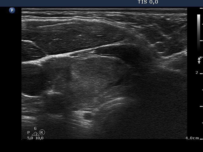
Right lobe, horizontal scan. Compared with the previous examination the lesion seems to be less well demarcated.