
Other edifying cases - Case 4. (ultrasonographic picture 1)
A muscle fiber mimicking a lobe in Hashimoto's thyroiditis

|
|
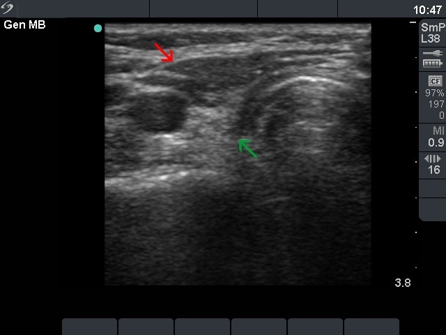
Right lobe, horizontal scan. In this view, it is equivocal which the lobe is. Comparing with the next images, it is clear that the hyperechogenic, fibrotic mass (green arrow) is the atrophic lobe, while ventral to the lobe there is a muscle fibre (red arrow).