
Other edifying cases - Case 4. (ultrasonographic picture 5)
A muscle fiber mimicking a lobe in Hashimoto's thyroiditis
|
|

|
|
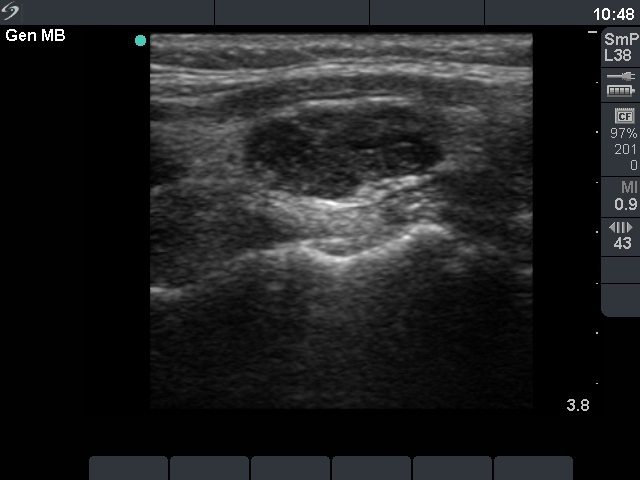
Left lobe, longitudinal view can be seen. A hypoechogenic lesion in the central part of the lobe is shown. This lesion seems like a nodule. However, in this case like in great part of Hashimoto's thyroiditis cases, this is not a nodule but only that part of the lobe where the thyroiditis is active. The echonormal parts above and under the hypoechogenic lesions are similar to the fibrotic-atrophic pattern demonstrated previously in the right lobe. Naturally, in such cases FNAC is indicated.