
|
|
The role of complex diagnosis - follicular proliferation - Case 4.
|
|


Clinical presentation: a 14-year-old boy was referred for an evaluation of a nodule discovered by himself.
Palpation: a hard, not freely moveable nodule in the isthmic part of the left lobe.
Hormonal examination: indicated euthyroidism with TSH 2.09 mIU/L.
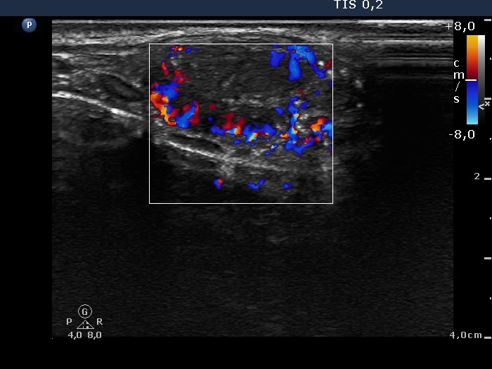
Ultrasonography revealed a hypoechogenic nodule in the ventromedial part of the left lobe. The nodule had irregular borders, contained microcalcifications and displayed perinodular and irregular intranodular blood flow.
FNAC report: follicular tumor.
Combining the US and FNAC results the probability of cancer was greater than the average.
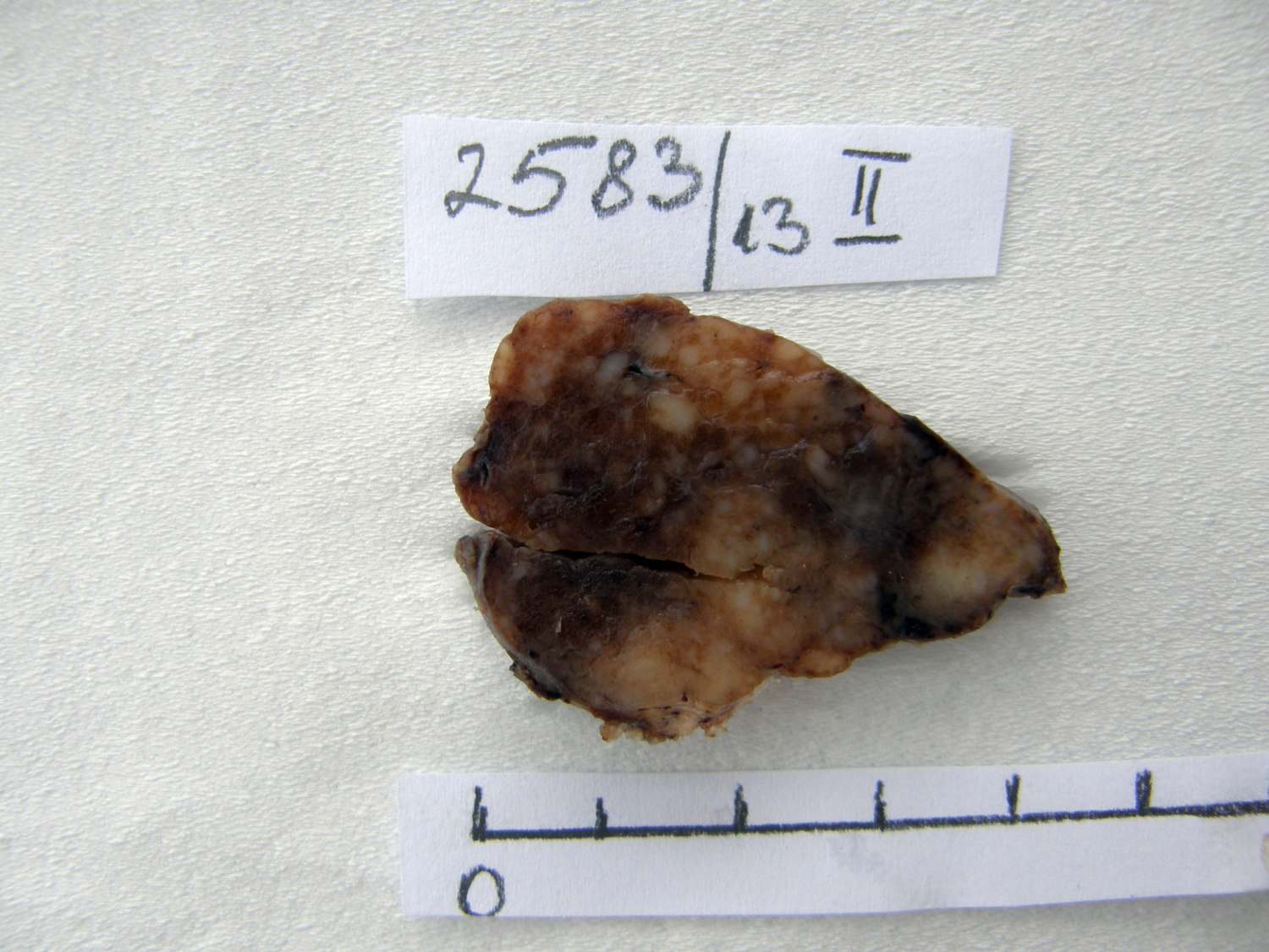
Histopathology: disclosed an embryonal-type follicular adenoma in the left lobe. Almost the entire lobe was consisted of the adenoma. There were within the nodule large fields of lymphocytic infiltration. In the middle portion of the nodule a papillary carcinoma was found with a maximal diameter of 15 mm with metastasis to 3 of the removed 9 lymph nodes in the left side of the neck.
Comment.
-
It is evident that the cytological material was gained not from the papillary carcinoma but from the adenoma.
-
It is hard to decide which portion of the lesion corresponds to the carcinoma.