
|
|
The role of complex diagnosis - follicular proliferation - Case 5.
|
|


Clinical presentation: a 47-year-old woman was referred for an evaluation of a nodule discovered on screening. Her daughter was operated on papillary carcinoma for several years.
Palpation: a not firm nodule in the right lobe.
Hormonal examination: indicated euthyroidism with TSH 1.34 mIU/L.
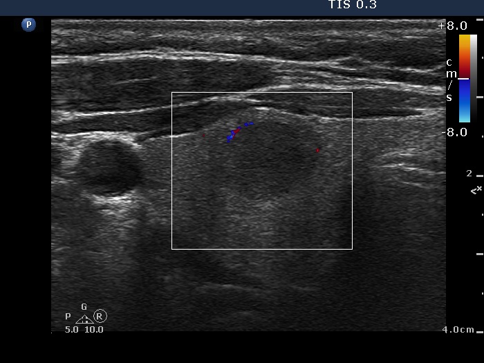
Ultrasonography revealed a moderately hypoechogenic nodule in the ventromedial part of the right lobe. The nodule presented neither perinodular blood flow nor halo sign.
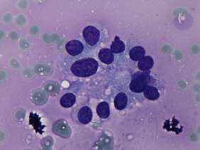
Cytology yielded a follicular pattern with predominance of microfollicles.
A combined ultrasound-cytological diagnosis was benign follicular proliferation.
Regular ultrasound examination was offered.
Comment. Although the presence of colloid stands against a follicular tumor, the predominance of isolated microfollicles suggests this opportunity. Considering the lack of halo and perinodular blood flow, the risk of a follicular tumor is very low.
.