
The role of complex diagnosis- follicular proliferation - Case 5. (ultrasonographic picture 3)
|
|

|
|
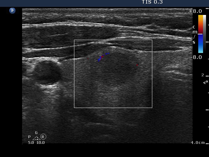
Right lobe, horizontal view, color Doppler mode. Although there are signs of a perinodular blood flow, this pattern belongs to the type 1 vascular group.