
Intranodular hyperechogenic figures - case 367 (ultrasonographic picture 5)
|
|

|
|
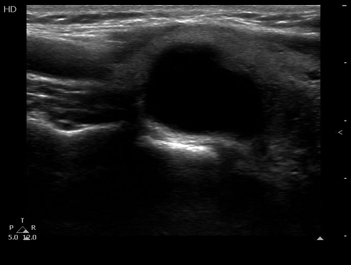
Right lobe, longitudinal view. Lower to the cystic part (right in the image) there is a smaller hypoechogenic solid area.