
|
|
The role of complex diagnosis - oxyphilic lesions - Case 4.
|
|


Clinical presentation: a 40-year-old woman requested a second opinion. She was treated for hypothyroidism for 7 years. She was examined because of her infertility. No thyroid ultrasonography was performed, yet.
Palpation: both thyroids were firm on palpation. The right lobe was enlarged and nodular on palpation.
Functional state: euthyroidism on daily 75 microgram levo-tiroxin (TSH 1.09 mIU/L).
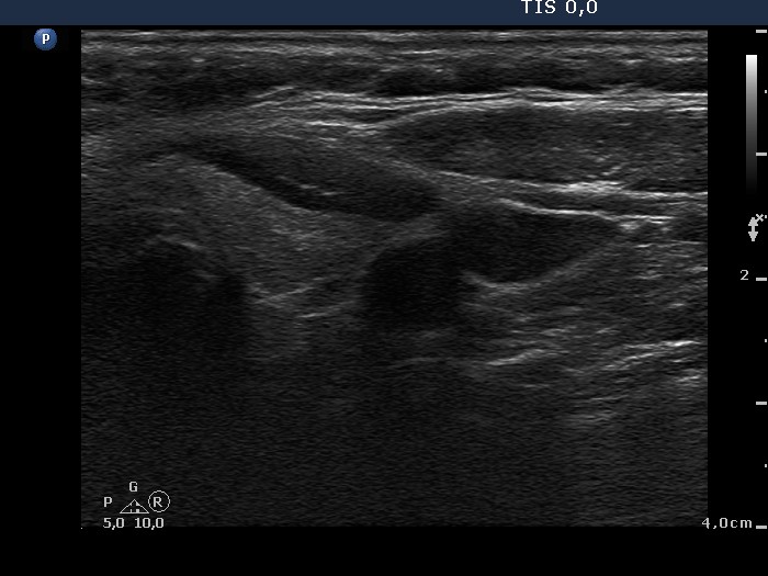
Ultrasonography: the thyroids were echonormal. There was a hypoechogenic nodule in the right lobe. The nodule displayed a halo sign and perinodular blood flow. There were smaller moderately hypoechogenic areas within both lobes.
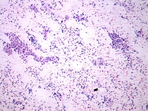
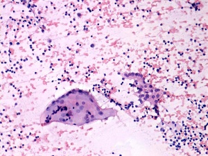
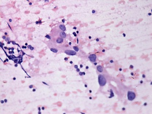
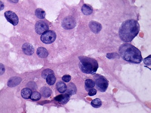
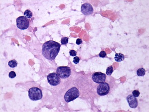
Combined cytological-clinical-sonographic diagnosis: Hashimoto's thyroiditis and Hürthle-cell tumor.
Histopathology disclosed Hashimoto's thyroiditis and Hürthle-cell adenoma.
Comments:
-
To perform ultrasonography is mandatory in each thyroid patient.
-
Although follicular cells display atypia and prominent nucleoli, the cytological picture itself is not suspicious. Taking the sonographic pattern into account the patient must have had a follicular-type tumor with great probability.