
|
|
Rare forms of thyroiditis - Case 9: Riedel's thyroditis
|
|


Clinical presentation: a 54-year-old woman was presented with a rapidly growing thyroid. The goiter had evolved over several weeks.
Palpation: both thyroids were enlarged, painless and very hard.
Functional state: hypothyroidism (TSH-level 72.0 mIU/L).
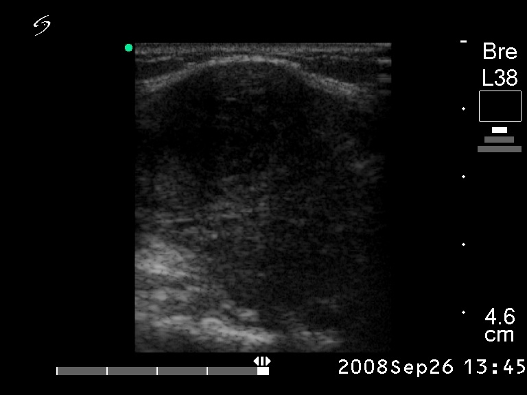
Ultrasonography: the whole thyroid was hypoechogenic. The extent of hypoechogenicity varied - in several parts of the thyroid was close to that observed in cystic lesions. The circumscribed lesions did not fit nodule. The vascularization was decreased.
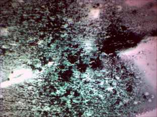
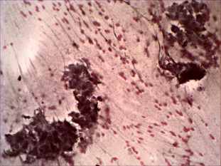
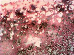
Cytological picture: there is no colloid in the background. High cellularity can be observed. The smear consists of compact clusters of round, as well as irregularly shaped cells, which show abundant syncytial cytoplasm and nuclear crowding. A few lymphocytes area in the background.
Combined cytological-clinical diagnosis: suspicion of MALT lymphoma.
Histopathology: Riedel thyroiditis which had destructed almost the whole thyroid. In the few islets of the parenchyma not influenced by fibrous thyroiditis, lymphocytic thyroiditis could be detected.