
Intranodular hyperechogenic figures - case 41
Follow-up examination 2 years later (ultrasonographic picture 1)
|
|

|
|
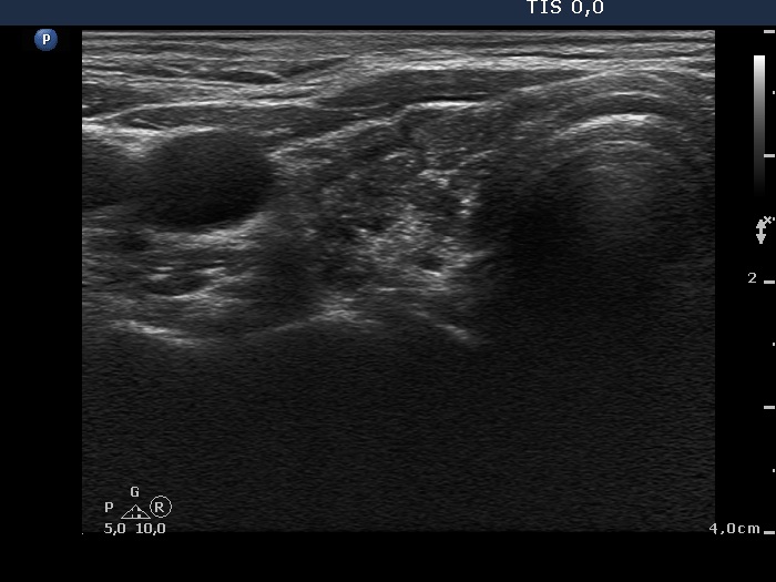
Right lobe, horizontal scan. A hypoechogenic, inhomogeneous lobe with pronounced fibrosis.