
Intranodular hyperechogenic figures - case 41
Follow-up examination 2 years later (ultrasonographic picture 6)
|
|

|
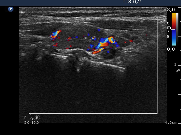
Left lobe, horizontal scan, color Doppler mode. The intranodular blood flow is a bit increased while the presence of a perinodular blood flow is doubtful.