
|
|
Intranodular hyperechogenic figures - case 420
|
|


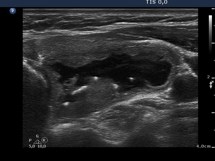
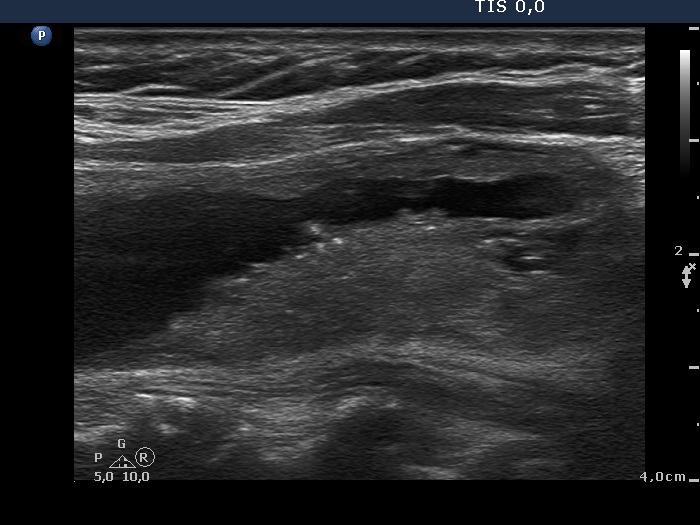


Clinical presentation: a 63-year-old woman requested an examination because of thyroid enlargement. She was operated on a multinodular goiter 20 years ago. She noticed a slow increase in the size of the right while a sudden increase in the left thyroid.
Palpation: a firm nodule in the right and an elastic one in the left lobe.
Functional state: euthyroidism (TSH 0.51 mIU/L, FT4 13.6 pM/L).
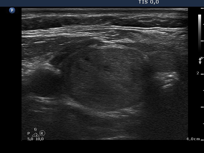
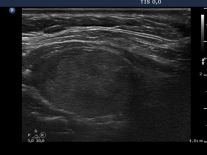
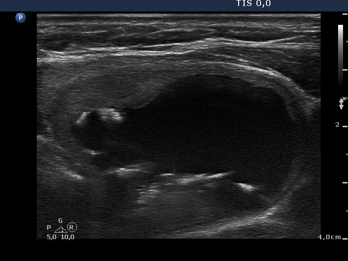
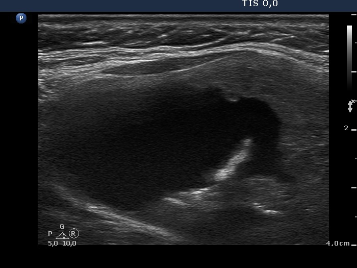
Ultrasonography. Almost the entire right lobe was composed of a minimally hypoechogenic nodule presenting perinodular blood flow. There was a central-type cyst in the left lobe with moderately hypoechogenic solid part. Both the solid and the cystic areas contained large and thick hyperechogenic lines and granules.
Cytology was performed form both lesions and resulted in colloid goiter and cystic lesion, right and left nodule, respectively. We removed 20 mL yellow fluid from the left lesion.
Comment. The intranodular hyperechogenic figures are unusually large. The ventral ones correspond correspond to thickened connective tissue and/or to large aggregates of colloid crystals (comet-tail artifacts) while those located in the back wall of the cyst are caused by posterior acoustic enhancement.
.