
The role of complex diagnosis - other examples - Case 5. (ultrasonographic picture 2)
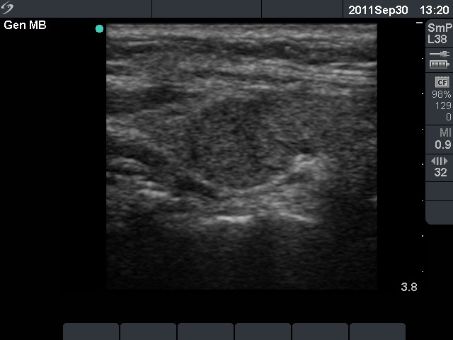 Right lobe, longitudinal scan. The upper and lower thirds of the lobe are less hypoechogenic compared with the middle third. Although this pattern suggests a moderately hypoechogenic nodule, the lesion is not regularly geometrical. |
