
|
|
The role of complex diagnosis - other examples - Case 5.
|
|


Clinical presentation: a 66-year old man was referred for aspiration cytology. Ha was operated on a multinodular goiter for 17 years. On ultrasound examination multiple nodules were described.
Palpation: no abnormality.
Hormonal examination: euthyroidism on daily 125 microgram levo-tiroxin.
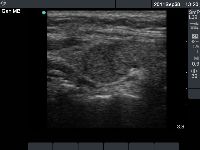
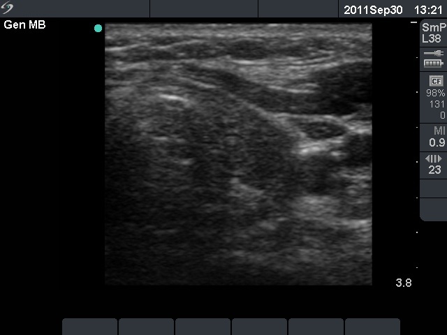
Ultrasonography: revealed hypoechogenic lobes surrounded with an echonormal rim. There were several discrete lesions, none of the fit to a nodule in a pathological sense.
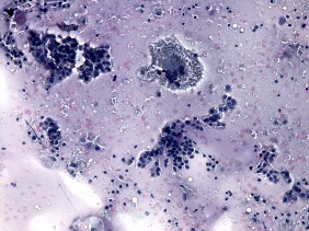
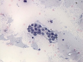
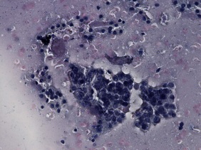
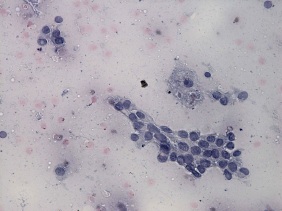
Cytology was performed from the hypoechogenic part of the right lobe. There were two cell populations on the smear including groups of atypical enlarged cells presenting groove and inclusiuon. Our cytological diagnosis was the following: papillary carcinoma cannot be excluded. We mentioned on our report that "although the cytological pattern is itself suspicious, considering the ultrasound presentation the risk of a papillary carcinoma is very low.
I discussed the diagnostic findings with the surgeon who managed the patient. I told him that although I had no other option than to raise the suspicion of papillary carcrinoma, I mean that the patient did not harbors carcinoma. The surgeon told me that on this FNAC report he has no option but to perform surgery.
Histopathology: benign, regular thyroid tissue without any discrete lesions.
Comments.
-
This case study was a turning-point in my practice. Never again I gave a distinct cytologcial diagnosis in patients whose cytological pattern was itself suspicious but the ultrasound pattern excluded the possibility of malignancy. In such situations I give a common ultrasound-cytological or a clinical-ultrasound-cytological diagnosis.
-
The cytological pattern meets the criteria of raising the suspicion of papillary carcinoma: there are atypical cells on the smear containing groove, occasionally inclusion.
-
It is worth to analyze the ultrasound: a central hypoechogenic area is surrounded with an econormal rim. Although the ultrasound presentation may be misleading by mimicking a large hypoechogenic nodule, this is one of the characteristic appearances of an intact, operated thyroid.