
The role of complex diagnosis - other examples - Case 5. (ultrasonographic picture 5)
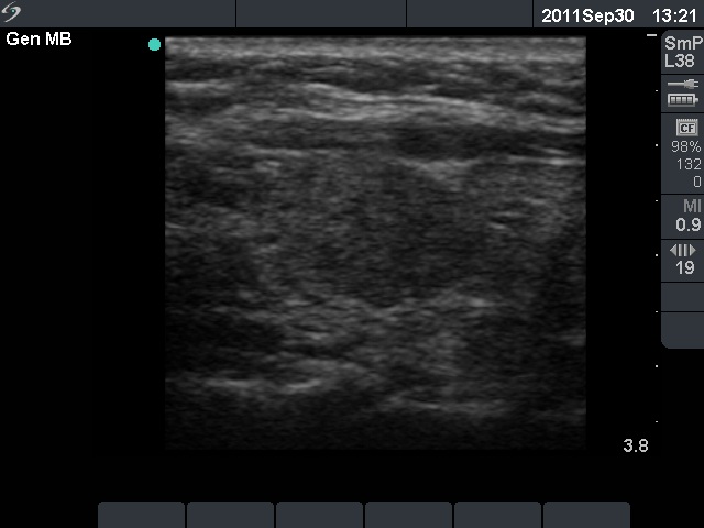 Left lobe, longitudinal scan. There is a larger, more hypoechogenic part in the middle portion of the lobe. Note the ventral echonormal incision which makes the impression that the lobe contained a larger moderately and a smaller minimally hypoechogenic nodule. |


