
Follicular adenoma - Case 16. (ultrasonographic picture 2)

|

|

|
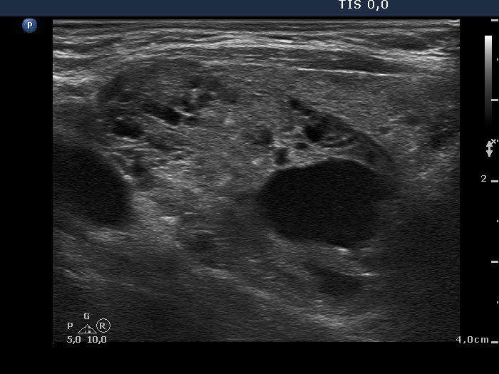
Right lobe, longitudinal scan. The nodule has sharp borders. There is a smaller lesion under and ventral to the large nodule (right in the image).


