
Subacute granulomatous de Quervain's thyroiditis - Case 20. (ultrasonographic picture 1)

|
|
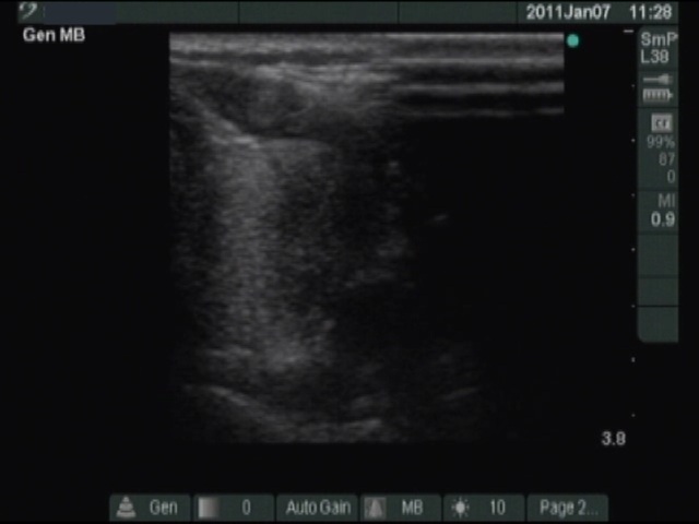
Left lobe, horizontal scan. A hypoechogenic lesion with blurred border is demonstrated here. This pattern may correspond both to papillary cancer and subacute de Quervain's thyroiditis.