
The role of complex diagnosis - follicular proliferation - Case 1. (ultrasonographic picture 3)
|
|

|
|
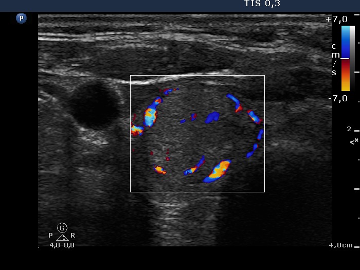
Right lobe, horizontal scan, color Doppler mode. A mixed type 2 and type 3 vascular pattern, i.e. the nodule presents both perinodular and intranodular blood flow.