
Differentiation of cystic thyroid lesions - Case 590 (ultrasonographic picture 3)
doi: 10.24390/thyrocase590.03

|

|

|
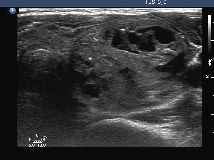
Left lobe, horizontal scan. A large cystic nodule occupies the nodule. The lesion has halo and presents intranodular hyperechogenic figures. The brightest one in the medial part of the nodule might cause differential diagnostic problem. It fully corresponds a microcalcification. However, in cystic lesion such figure is more probably also caused by back wall cystic enhancement.


