
Differentiation of cystic thyroid lesions - Case 6 (ultrasonographic picture 5)
doi: 10.24390/thyrocase6.05
|
|

|
|
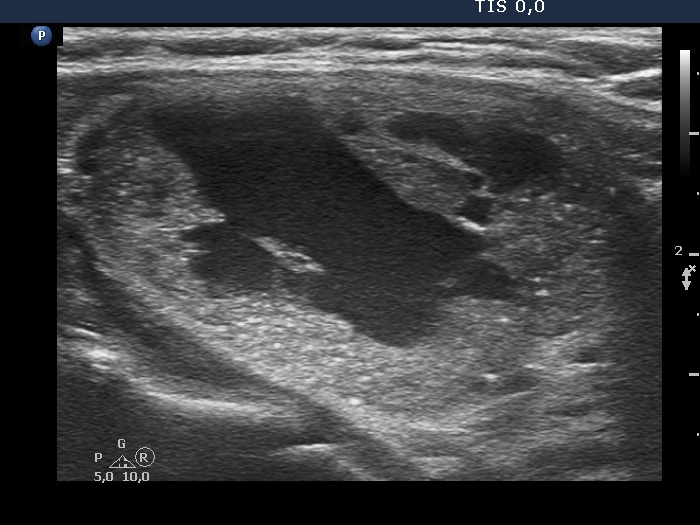
Left lobe, another longitudinal scan. Beside echogenic figures caused by posterior back wall enhancement there are other echogenic granules the origin of which is doubtful. Nonetheless, these probably belong also to the back wall figure subgroup and not to the punctate echogenic foci (microcalcifications) category.