
Differentiation of cystic thyroid lesions - Case 6 (ultrasonographic picture 9)
doi: 10.24390/thyrocase6.09

|

|

|
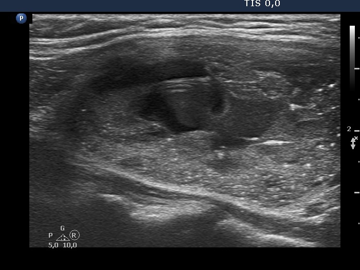
Left lobe, longitudinal scan - after removing 8 mL bloody cystic fluid. The image was recorded 30 seconds later than the former image. The cloudy mass in the central part of the nodule corresponds to the blood refilled just after the aspiration.


