
|
|
Differentiation of cystic thyroidlesions - Case 636doi: 10.24390/thyrocase636.00
|

|


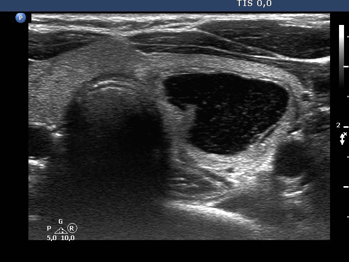
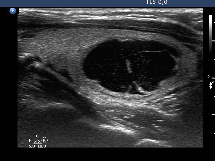

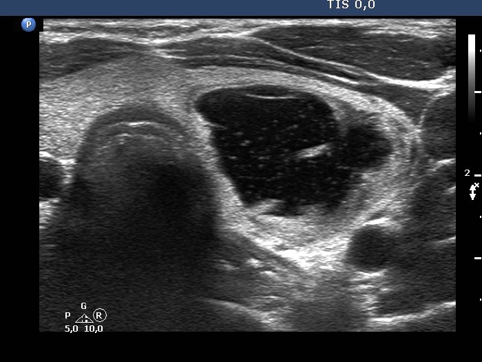
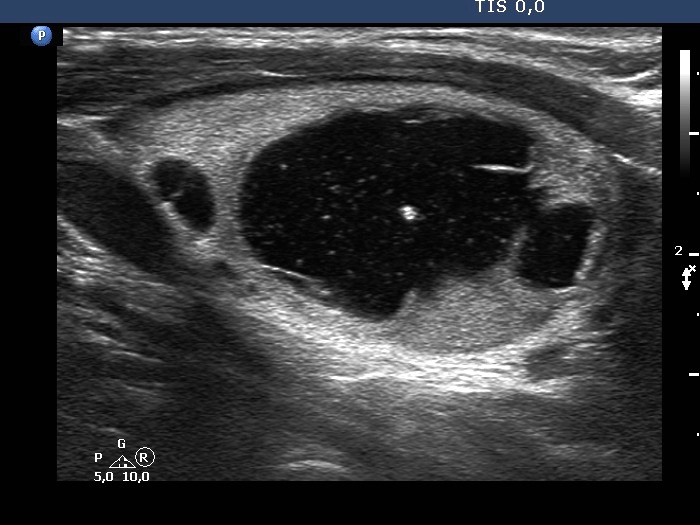
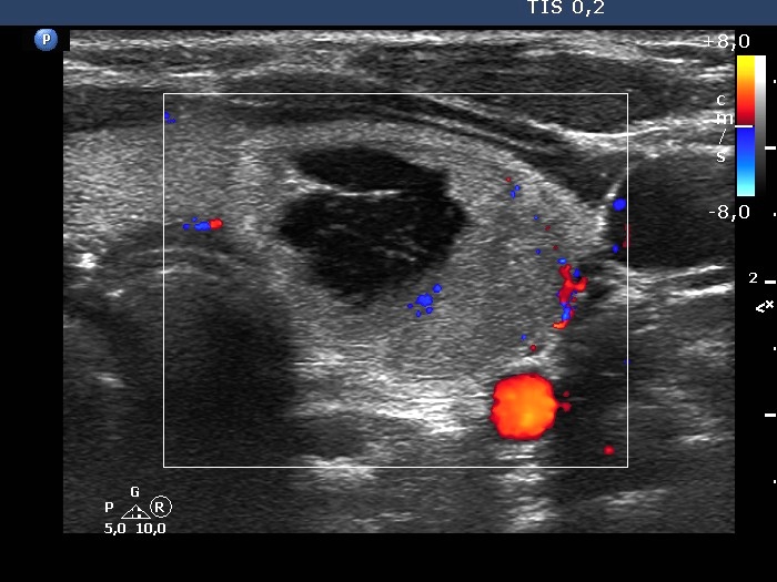
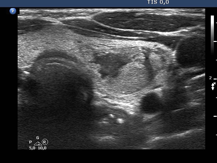
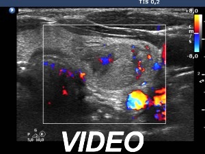
First examination (first row of images)
Clinical presentation. A 36-year-old woman noticed enlargement of the left thyroid after an upper airway infection.
Palpation. The left lobe had an elastic nodule.
Result of blood tests: TSH 1.69 mIU/L.
Ultrasonography. The thyroid was echonormal. There was a central-type cystic lesion in the left lobe. The cystic part presented numerous echogenic granules without a tail.We aspirated 5 mL bloody fluid. Aspiration cytology resulted in non-diagnostic cystic lesion.
Suggestion: thyroid ultrasound in a year.
Second examination one year later (second row of images)
Clinical presentation. The patient had no compaints.
Palpation: unchanged.
Result of blood tests: TSH 2.44 mIU/L.
Ultrasonography. The cystic nodule had refilled. The presentation remained the same.7 mL brown fluid was aspirated. Cytology: benign, cystic lesion.
Suggestion: ultrasound in two years.


