
Differentiation of cystic thyroidlesions - Case 636
One year later (ultrasonographic picture 9)
doi: 10.24390/thyrocase636.2.09

|

|

|
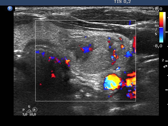
Left lobe, horizontal scan, color Doppler mode - after aspirating 7 mL brown fluid. The nodule presents signs of perilesional blood flow.


