
|
|
Follicular carcinoma - Case 2.doi: 10.24390/thyrocase68.00
|
|


Clinical presentation: A 29-year-old woman was referred for an evaluation of a nodule discovered by her GP on evaluation of a viral infection.
Palpation: The left lobe was enlarged and had a large moderately firm nodule.
Hormonal examination indicated euthyroidism with TSH 3.73 mIU/L. The aTPO level was 2.3 U/mL.
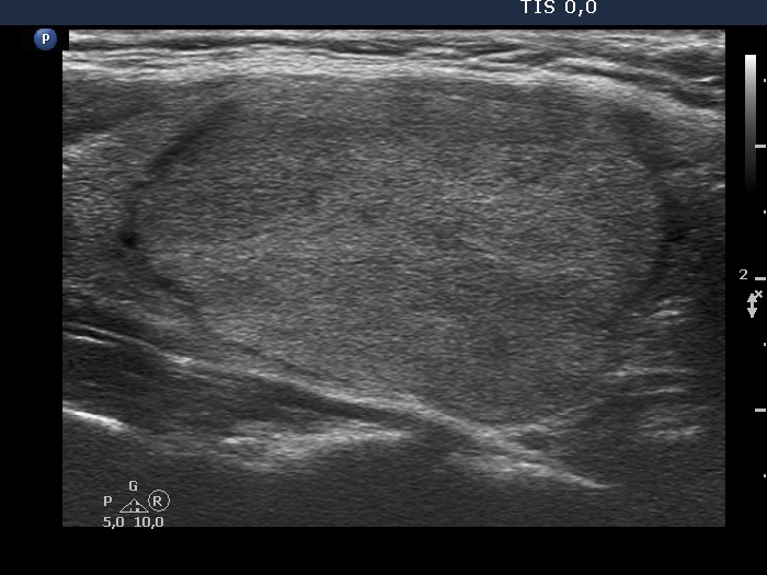
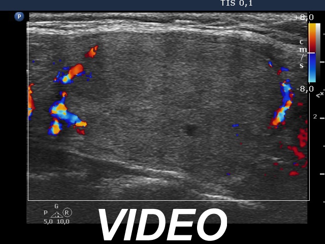
Ultrasonography. The thyroid was echonormal. There was a large nodule in the left lobe with the dimensions of 32, 25 and 42 mm, width, depth and lenght, respectively. The nodule was echonormal and has minimally hypoechogenic fields. The lesion had partly lobulated margins and presented halo sign and perinodular blood flow.
Cytology resulted in follicular proliferation.
A combined ultrasound-cytological diagnosis resulted in follicular tumor.
Histopathology disclosed follicular cancer with both capsular and vascular invasion.
.