
|
|
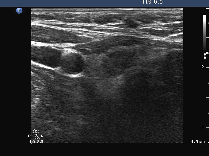
Follicular carcinoma - Case 1.
|
|


Clinical presentation: a 54-year-old man was referred for an evaluation of a nodule discovered by himself. The patient suffered from hoarseness for two months.
Palpation: a hard nodule in the left lobe.
Hormonal examination indicated euthyroidism with TSH 0.87 mIU/L.
Ultrasonography: there was a small hypoechogenic nodule in the right, while a large one in the left lobe. The left nodule was deeply hypoechogenic, had irregular, blurred borders. This lesion did not exhibit perinodular or intranodular blood flow.
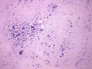
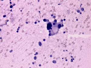
FNAC was repeatedly not diagnostic, we gained only bloody-necrotic material. Cytological images of the intraoperative imprint smear are presented.
Measurement of thyroglobulin in the wash-out of the needle was performed and resulted in 23 ng/ml, a very high level. Therefore the possibility of a metastasis could be excluded. The clinical and ultrasonographic picture was more than suspicious, therefore the patient were sent to surgery.
On further investigation palsy of the left recurrent nerve was established. CT scan disclosed multiple metastatic foci in the lung.
Histopathology: disclosed widely invasive follicular cancer in the left lobe.
.