
|
|
Medullary carcinoma - Case 10.
|
|


Clinical presentation: a 68-year-old woman with a nodule discovered 2 months earlier. Nodules were palpable in both lobes. The nodule in the left lobe was hard on palpation.
Functional state: euthyroidism with TSH-level 0.83 mIU/L.
Ultrasonography: a large hypoechogenic nodule in the left lobe with hyperechogenic patches without acoustic shadow Therefore these are neither signs of coarse calcification nor microcalcification. These correspond to amyloid deposits.
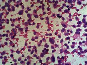
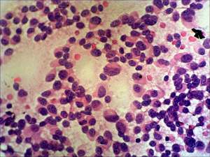
Cytology. Extremely cellular picture. The cells form neither follicles nor papillary fronds, they are arranged in loose syncytial structures and dissociated. The atypical cells are polygonal with eccentric nuclei. Note triangular forms specific for medullary cancer.
Cytological diagnosis: medullary cancer.
Histopathology: medullary cancer with metastasis to the lymph node of the neck.