
Oxyphilic adenoma - Case 2. (ultrasonographic picture 3)
|
|

|
|
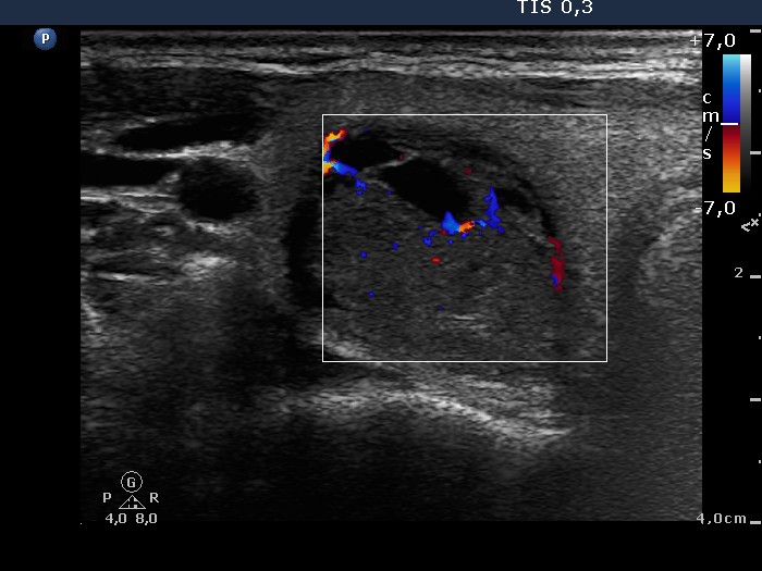
Right lobe, horizontal scan, color Doppler mode. Combined type 2 and type 3 vascular pattern, i.e. both intranodular and perinodular blood flow can be visualized.