
Subacute granulomatous de Quervain's thyroiditis - Case 35.
Follow-up examination 3 weeks later (ultrasonographic picture 5)
|
|

|
|
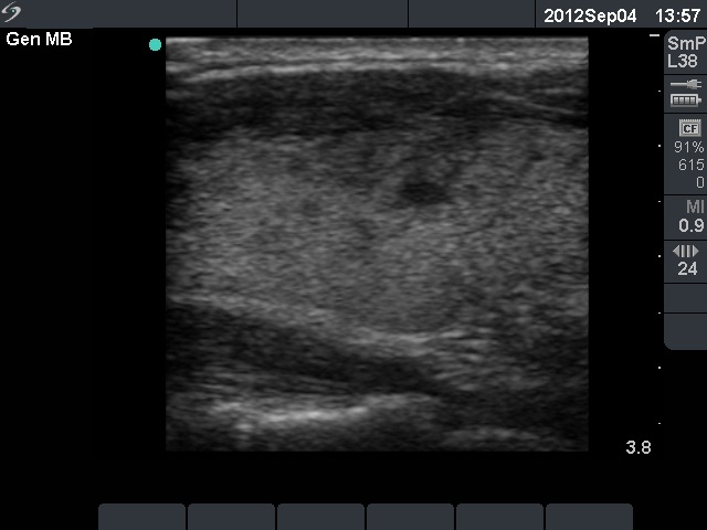
Left lobe, longitudinal scan. There is a hypoechogenic area in the ventral part of the lobe. Although the lesion has decreased in size, the borders are blurred.