
|
|
Oxyphilic adenoma - Case 22.
|

|


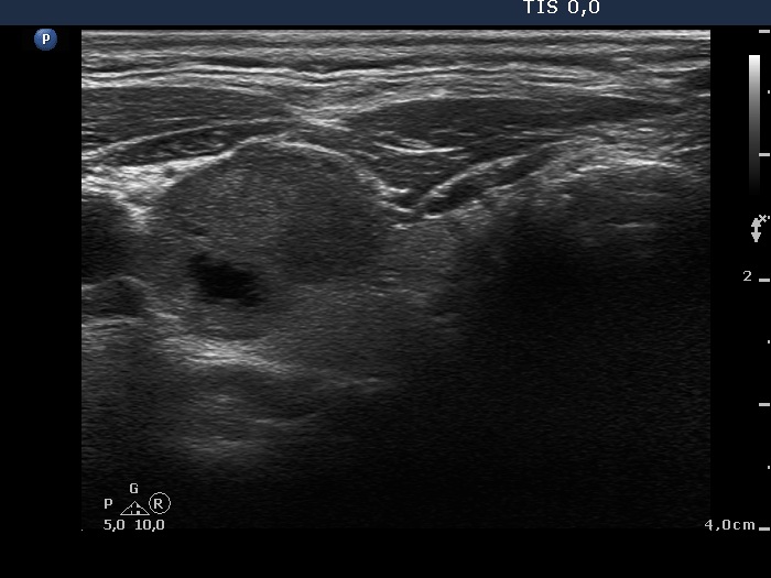
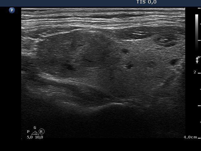
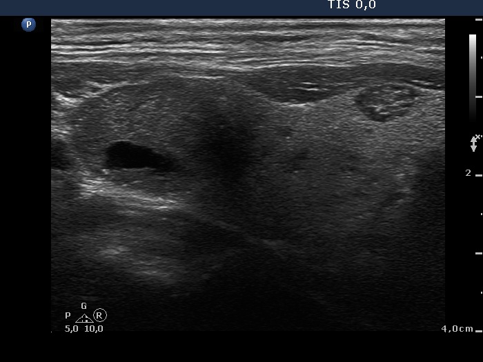
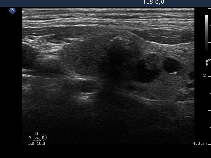
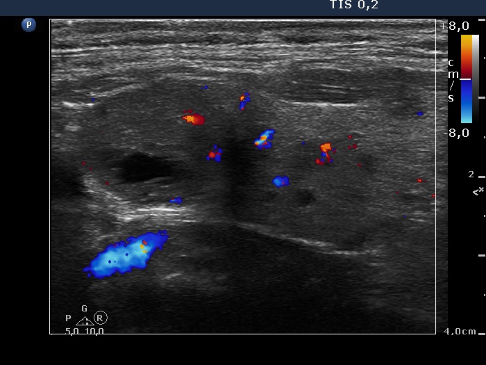
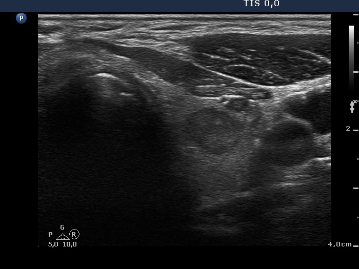
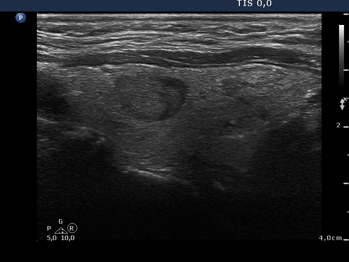
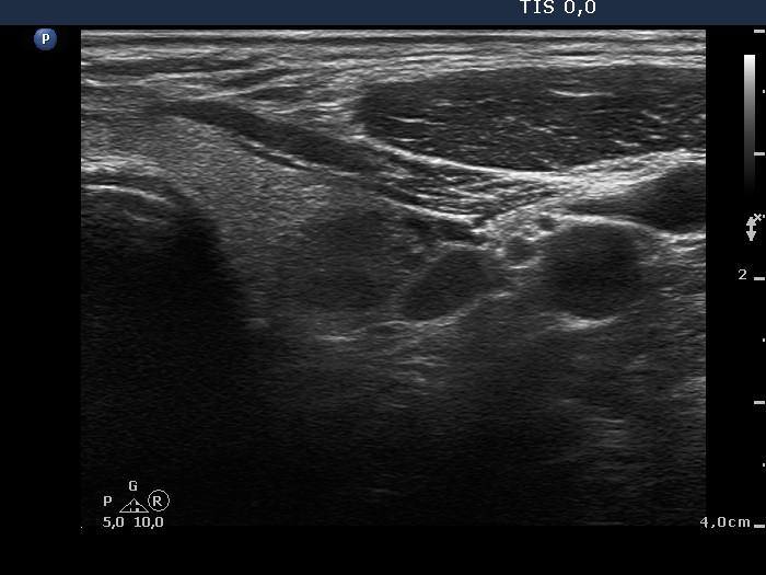
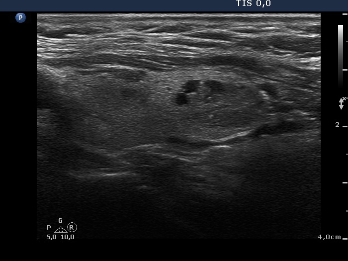
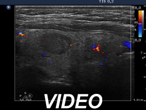
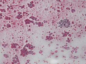
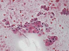
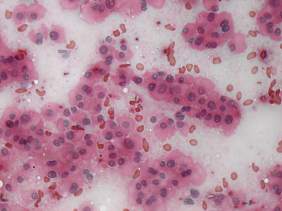
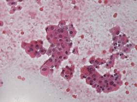
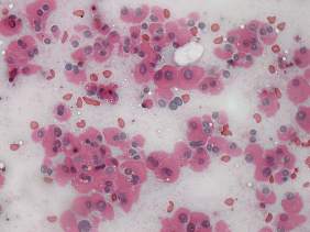
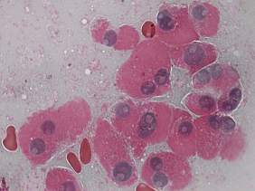
Clinical presentation: a 74-year-old man was referred for evaluation of thyroid nodule discovered on PET CT scan which was performed because of herpes zooster.
Palpation: left lobe contained multiple, moderately firm nodules.
Functional state: euthyroidism (TSH 1.63 mIU/L).
Ultrasonography revealed a multinodular goiter with lesions of different echogenicity. Three nodules were aspirated. The lesion in quetion was located in the upper part of the left lobe.
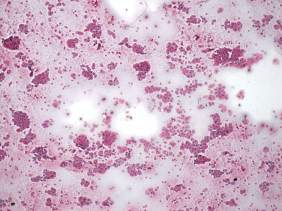
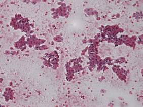
Aspiration cytology resulted in oxyphilic tumor.
Histopathology disclosed oxyphilic cell adenoma.
The lesion in question is located in the the left lobe.
Comments. The diagnostic values of nuclear inclusions is limited in the event of an oxyphilic lesion. Moreover, the extreme proportion of this figures should itself raise concern about the origin of these figures. In contrast with drying artifacts, the inclusions in this case have regular borders and show a dark sharp rim. The cause for the difference in histopathological and cytological is not known.


