
Oxyphilic adenoma - Case 22. (ultrasonographic picture 9)

|

|

|
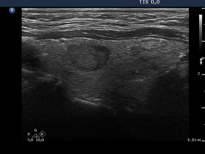
Upper part of the left lobe, longitudinal view. The cytological images come from the upper moderately hypoechogenic nodule (left in the image).


