
|
|
Chronic lymphocytic thyroiditis - Case 77.
|

|


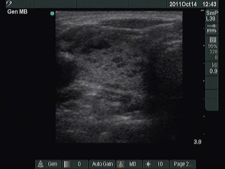




First examination on 16th gestational week (first row):
Clinical presentation: a 37-year-old woman was referred to a follow-up evaluation of Hashimoto's thyroiditis.
Palpation: both thyroids were nodular and firm on palpation.
Hormonal investigation indicated euthyroidism with TSH-level 1.87 mIU/L.
Ultrasonography: the right thyroid was echonormal with around 15% of the hypoechogenic areas. The left lobe contained larger hypoechogenic-hyperechogenic circumscribed areas which did not fit a nodule in a pathological sense.Cytology was performed after the wish of the patient and resulted in Hashimoto's thyroiditis.
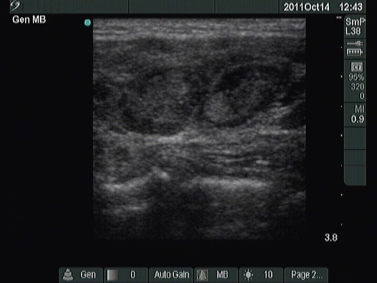
Follow-up examination 7 weeks after delivery (second row):
Clinical presentation: the patient had no complaints.
Palpation: both thyroids were nodular and firm on palpation.
Hormonal investigation indicated euthyroidism with TSH-level 1.97 mIU/L.
Ultrasonography: the sonographic pattern was unchanged.Suggestion: TSH-determnation every year, ultrasound examination every 3 years.


