Benign nodular hyperplasia - Case 2. (ultrasonographic picture 8)
doi: 10.24390/thyrocaseconp772.13
|
|

|
|
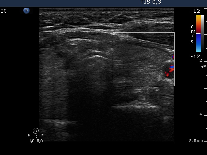
Left lobe, horizontal view, color Doppler mode. There is no vascularization within the lobe.

|
|
|
|
Left lobe, horizontal view, color Doppler mode. There is no vascularization within the lobe.