
Oxyphilic adenoma - Case 20. (ultrasonographic picture 4)

|

|

|
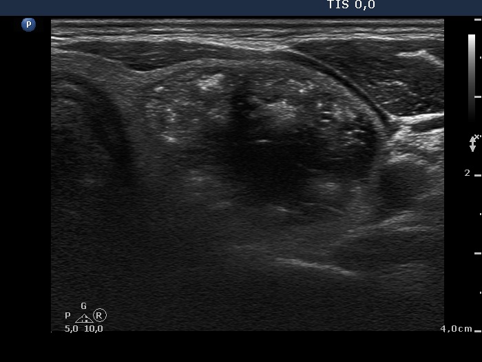
Left lobe, horizontal scan. There is a mixed nodule with an echonormal part presenting hyperechogenic figures, both granules and larger patches.


