
|
|
Oxyphilic adenoma - Case 20.
|

|


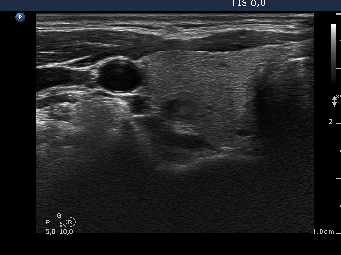
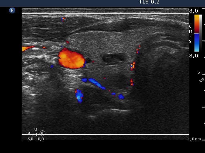
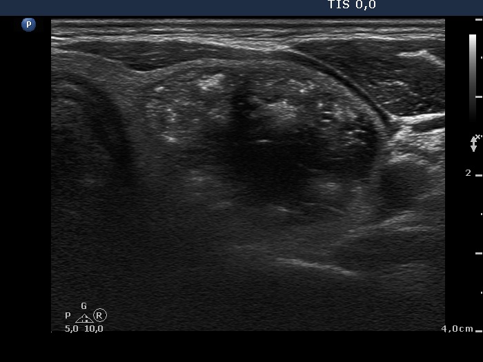
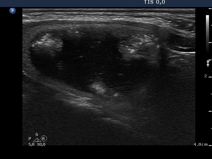

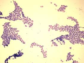
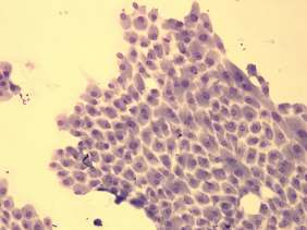
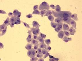
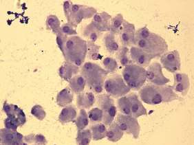
Clinical data: a 41-year-old woman was referred for an evaluation of a thyroid nodule detected on physical examination. She was investigated because of fatigue and hypertension.
Palpation: an elastic nodule in the left lobe.
Laboratory tests: TSH 0.81 mIU/L, anti-TPO 0.1 U/mL.
Ultrasound. The thyroid was echonormal. The right lobe presented several small hypoechogenic and cystic lesion. There was a large mixed nodule occupying almost the entire left lobe. The solid part of the lesion was echonormal and contained hyperechogenic granules and patches. The lesion presented a type 1 vascular pattern. Only a small part of the nodule was surrounded with halo.
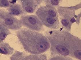
Two US-guided aspirations were performed. A minimal amount of brown fluid was gained. Preliminary cytological diagnosis: tumor with great probability an oxyphilic tumor.
Wash-out thyroglobulin level exceeded 478 microgram/L, serum calcitonin was calcitonin 0.84 pM/L (normal value: 0-3.36).
Final cytological diagnosis: oxyphilic tumor.
Left lobectomy was performed. Histopathology disclosed an oxyphilic adenoma
Comments.
-
The nodule is surrounded with halo but lacks perinodular blood flow. The latter is explained by the compression of the cystic fluid.
-
The solid part is very unusual. The presence of hyperechogenic patches raises the suspicion of a medullary carcinoma.


