
Intranodular hyperechogenic figures - case 808
Follow-up investigation 3 years after first visit (ultrasonographic picture 5)
doi: 10.24390/thyrocase808.2.05
|
|

|
|
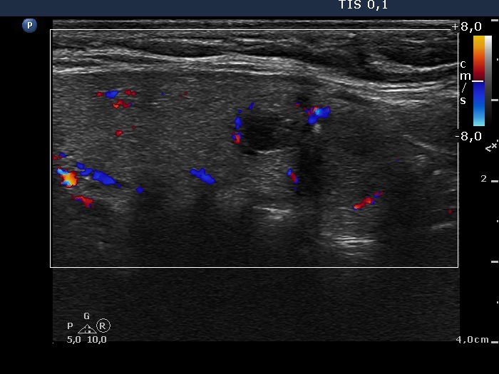
Right lobe, longitudinal scan, color Doppler mode. The nodule in the central-lower part of the lobe shows signs of perilesional blood flow.