
Intranodular hyperechogenic figures - case 808
Follow-up investigation 3 years after first visit (ultrasonographic picture 9)
doi: 10.24390/thyrocase808.2.09
|
|

|
|
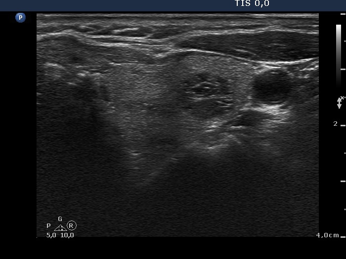
Left lobe, horizontal scan. The nodule in this lobe remained unchanged as regards the size and ultrasound presentation.